Focus of the Centre for Artificial Intelligence in Oncology
We focus on the applications of AI in those oncology-related tasks that are too time-consuming or difficult or even impossible to be performed by human. We try to contribute both to the general progress in the cognition of cancer-related phenomena and to the personalized patient care.
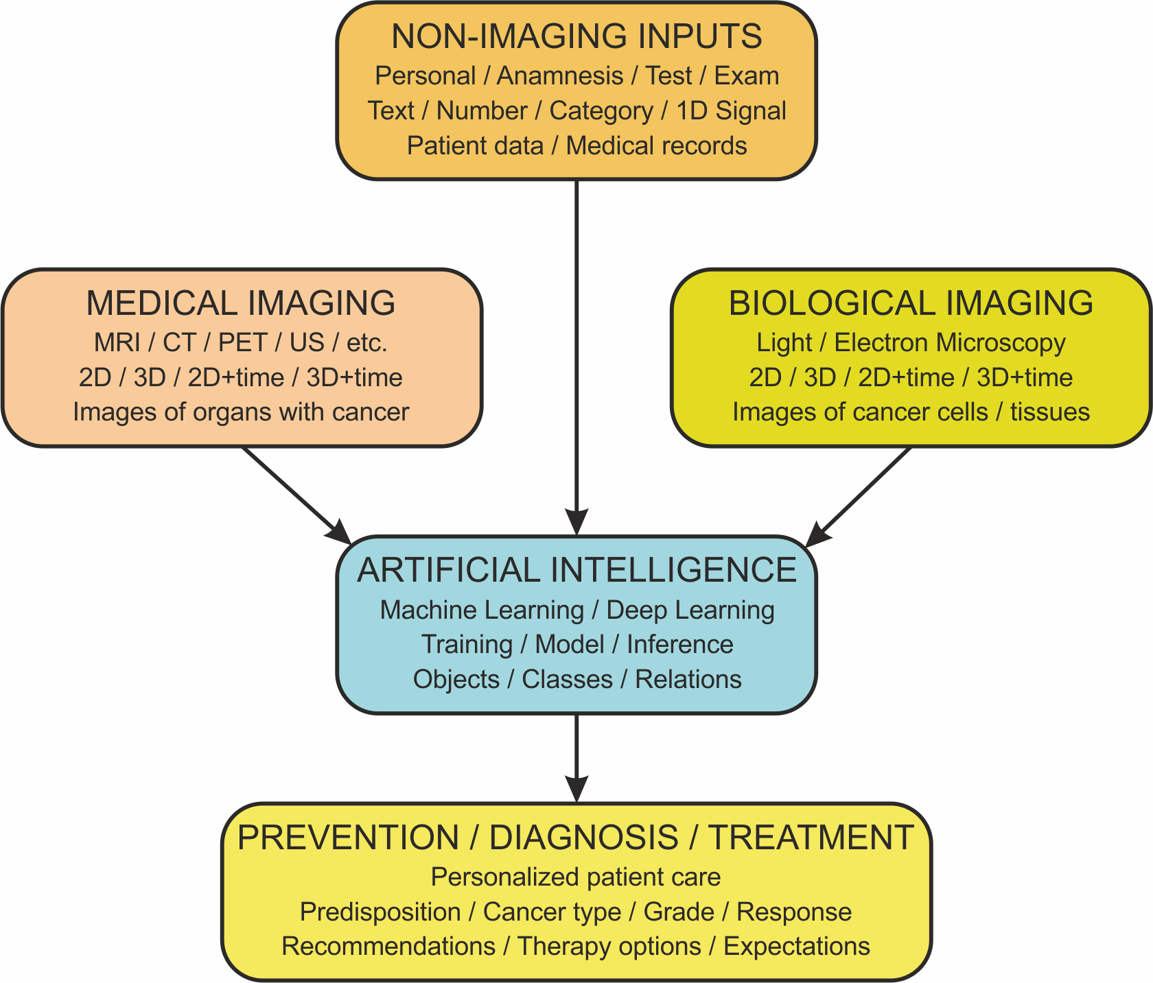
We work with three types of input data:
Medical imaging data
2D or 3D images or videos of an investigated organ of a patient with cancer acquired using magnetic resonance imaging (MRI), computed tomography (CT), positron emission tomography (PET), ultrasound (US) or other modalities
Biological imaging data
2D or 3D images or videos of cancer cells or tissues, including artificially grown spheroids or organoids after applying certain drug, acquired using light or electron microscopy
Non-imaging data
Patient data (e.g. physical, family or lifestyle characteristic + clinical notes and other medical records) integrated with related biomedical datasets (covering for instance genes, mutations, proteins, pathways, diseases, interventions, drugs or publications)
Artificial Intelligence
The input data are processed using AI, typically using machine learning, mostly deep learning approaches. Because supervised learning is mostly used, sufficient amount of data must be complemented with correct answers provided by experts (so-called gold-standard reference annotations) for the algorithm development phase. After training and validation, the developed method/model can be applied to further non-annotated data. The AI result should then be interpreted with care and combined with human intelligence.
For image data, we mostly perform the following tasks using AI:
- Image quality improvement such as de-noising, de-blurring or increasing resolution
- Synthesis of additional modalities saving instrument and patient time
- Detection of relevant objects or events of interest
- Segmentation of relevant objects assigning each pixel/voxel to some class/object
- Classification of objects or whole images/patches
- Tracking of objects in time
For non-image data (that may include image analysis results as well), we mostly attend to the following:
- Collecting, extracting, cleaning and preprocessing data from oncological patient records and public datasets of relevant biomedical entities (genes, mutations, proteins, pathways, diseases, interventions, drugs and toxic substances, etc.).
- Integrating the preprocessed data in uniform, semantically-interlinked resources (such as knowledge graphs or ontologies).
- Augmenting the integrated resources by automated inference (e.g., link prediction, rule- or logics-based reasoning or knowledge base completion).
- Developing predictive machine learning and explainable AI models on top of the collated oncological knowledge bases.
- Developing intuitive querying and visual exploration interfaces on top of the models in a co-design process with cancer biologists, pharmacologists, doctors, patients and clinical psychologists.
- Developing and evaluating AI-enabled decision support prototypes by their preliminary deployment in clinical settings and comparison with the current clinical practices.
- Exploring the opportunities for building predictive models on top of multimodal data (i.e., incorporating the imaging data into the models based on non-image data, or the other way around).
Finally, AI results serve clinicians as an aid for taking decisions concerning prevention, diagnosis or treatment either as general know-how to be taken into account or as a specific incentive for a particular patient.